The new British research is the first to reveal striking differences in areas of the brain based on scans taken before and after a coronavirus infection.
are associated with subtle tissue damage and accelerated losses in brain regions tied to the sense of smell, as well as a small loss in the brain’s overall volume, a new British study finds. Having mild COVID-19 is also associated with a cognitive function deficit.
The red-yellow regions are the parts of the brain that shrank the most in the 401 SARS-CoV-2 infected participants, compared with the 384 noninfected participants. Gwenaëlle Douaud, in collaboration with Anderson Winkler and Saad Jbabdi, University of Oxford and NIH.Monday in Nature, also stands out because the lion’s share of its participants apparently had mild COVID-19 — by far, the most common outcome of coronavirus infections.
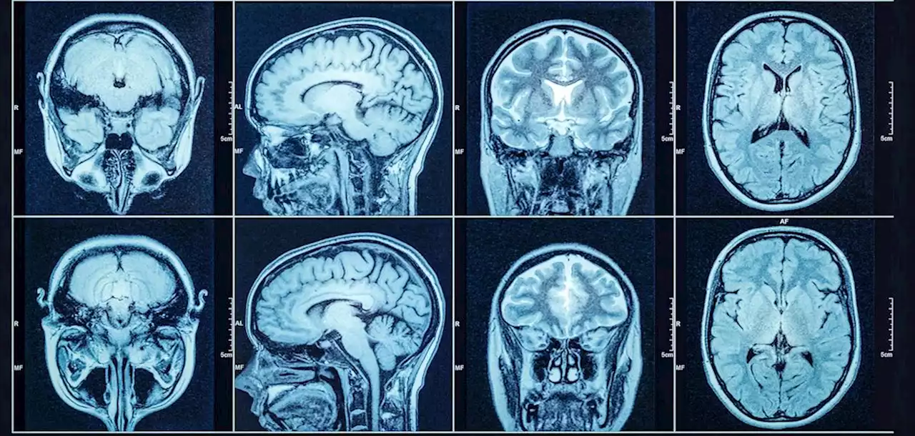
Between the pairs of MRIs, which were separated by an average of about three years, the researchers observed a striking trend among those who had COVID: a greater loss of what’s known as gray matter in the brain, as well as a higher rate of abnormalities in the brain tissue. Gray matter, which appears gray on certain brain scans, is comprised of various cells, including neurons.